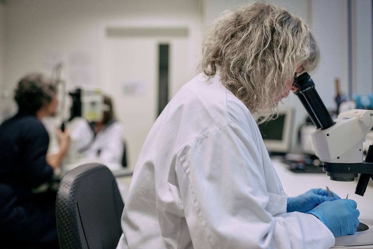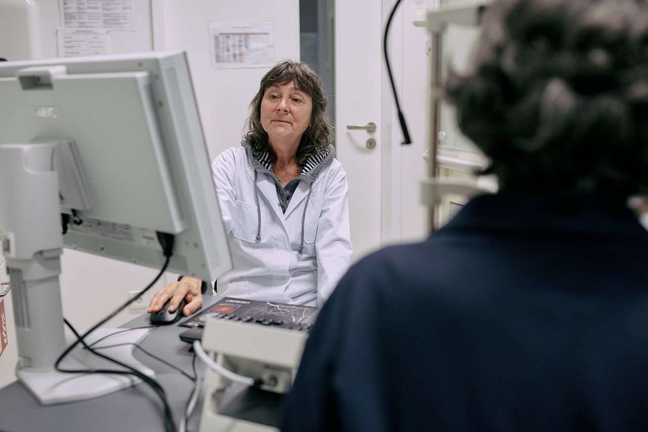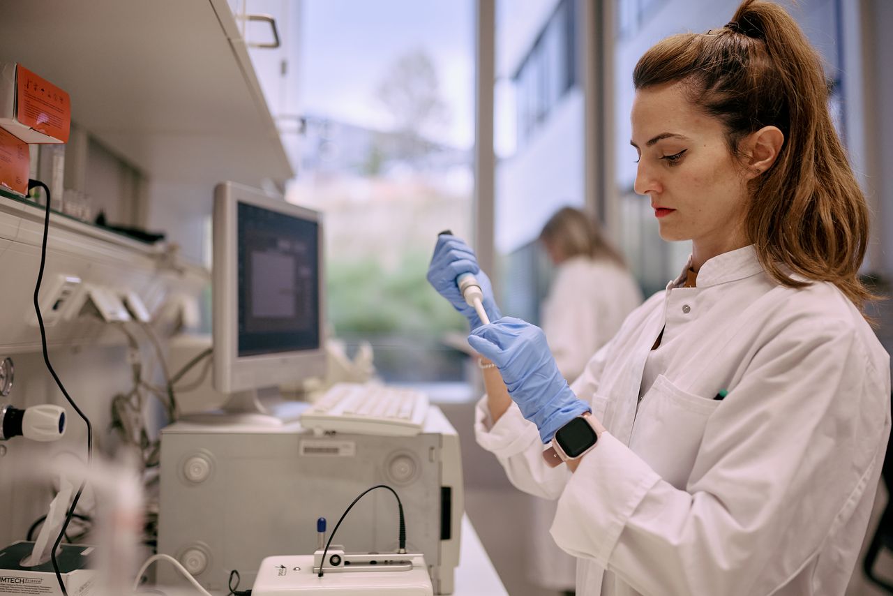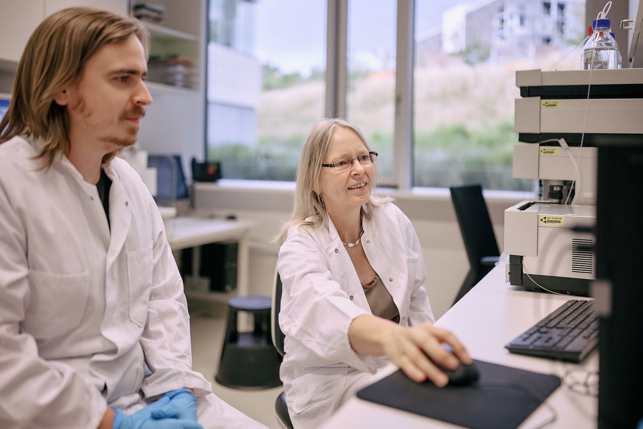
Neurobiology of the Eye
The Research Topics
Our central research goal is to gain a better understanding of how visual experience and genetic factors affect eye growth and the development of myopia. We are studying these questions in chicken and mouse models, but also in human subjects. When animal models wear spectacle lenses or diffusers in front of their eyes, they develop myopia (eye grows longer, with negative lenses and diffusers ) or hyperopia (eye remains shorter, with positive lenses). The predictable effect of these visual perturbations on eye growth permits to study the underlying retinal image processing, i.e. how defocus is detected and quantified over time. Furthermore, one can learn how the output of retinal image processing merges into the release of growth signals from the retina that reach the retinal pigment epithelium where they may be converted into other signals that pass through choroid to the sclera.
Myopia is a multi-layered problem since it develops as a complex interaction of genes and (mainly visual) environment. We study myopia at several levels:
- Optics & image processing: while experiments in animals clearly show that defocus, including its sign, is detected by the retina in a few minutes, it is still not clear how this is done. While experiments in chickens have shown that defocus is detected in all parts of the visual field to control eye growth locally, it is not clear how this mechanism interacts with accommodation. Accommodation must clearly change the pattern of defocus over time and needs to be taken into account. While humans have only a small central area with high visual acuity, the fovea, and large amounts of astigmatism in the periphery, experiments in primates have nevertheless shown that peripheral defocus provides the major input into the control of axial eye growth - the fovea seems not very important. Since it is known that accommodation is largely driven by the fovea, the interaction of peripheral defocus (which controls eye growth) and fovea (which controls accommodation) reaches a new level of complexity. It is necessary to measure the patterns of defocus over time with intact accommodation.
- Electrophysiological approach: using the microelectrode array, ganglion cell activity can be studied in isolated mouse or chicken retina when focused and defocused movies were projected on the retina.
- Pharmacological approaches and screening studies: Identifying retinal messengers that are released from the retina when defocus is imposed. This makes it possible to choose drugs to alter axial eye growth pharmacologically. In turn, intravitreal application of agents helps to identify transmitter systems that are involved in the signalling cascade. New candidates were also identified in microarray studies and proteom screening studies (in collaboration with Novartis 2006). More recently, we have found that insulin in the eye is a powerful stimulator of myopia. Conversely, glucagon was found to inhibit myopia development in the chicken. Immunhistochemical studies are used to localize the candidates to certain layers and cell types (i.e. glucagon: Mathis & Schaeffel, 2007; TGF-ß: Mathis & Schaeffel 2010). A promising approach is also to use OCT to measure choroidal thickness changes which are always precursors of the changes in axial eye growth – thicker choroid results in less growth, and is therefore desirable when myopia should be inhibited.
- Psychophysical studies: our major interest is in spatial adaptation, i.e. how the retina and the cortex change their spatial filters to improve vision in the presence of defocus, astigmatism and aberrations. After adaptation to astigmatism, visual acuity is clearly improved even when astigmatism persists but the adaptation mechanism is not understood. It is clear that the adaptation is selective for the axis of astigmatism. Also the time courses of adaptation are of interest since they provide information about the temporal integrator of the defocus information.
- Interaction of myopia development with bright light and the role of retinal dopamine: after it was found in children that the average amount of myopia is inversely correlated with the number of weekly hours spent outdoors, we have studied this in chickens wearing diffusers and found that they also develop only half as much myopia if they are kept outdoors. We found that this effect is mediated by the high level of ambient illuminance outdoors and that it is very likely that the increased release of dopamine from the retina plays an important role. To study time course and dose-response relationship, OCT measurements of choroidal thickness in bright light may provide important hints.
- Behavioral studies: we have developed automated procedures based on the optokinetic head nystagmus to measure contrast sensitivity and contrast adaptation in chickens and mice.
- Studies on the optical and adaptational limits of vision: this direction was initiated by a new initial training network (ITN) of the Madam Curie program (http://www.itn-opal.eu/). Currently, the origin and effects of transverse chromatic aberration are studied, as well the origin of the human foveal blue scotoma. New techniques are developed to measure both longitudinal and transverse chromatic aberration. We also develop new techniques to measure fixational eye movements and their interaction with the spatial content and luminance of the viewing target.
- Adaptation to vertical disparities: Spectacle lenses have always prismatic effects which result in a lateral displacement of the retinal images in both eyes with respect to each other. In particular vertical displacements are disturbing, and we are studying how eye movements and visual cortex deal with heterophorias. Furthermore, we are studying how high frequency random spatial jitter of random dot pattern to be fused affects stereopsis.
- Development of optical instruments: There is a long-standing history of development of instruments and new procedures to measure optical features of the eye. This started with the first generation PowerRefractor, an eccentric infrared photorefractor. Its current versions are developed and marketed by PlusOptix, Nuernberg, Germany (http://www.plusoptix.de). An overview showing some of the newly developed techniques can be found on a poster (link to poster). Some more descriptions are provided below (“Methodology and technology development”).
Foto: Beate Armbruster / © Universitätsklinikum Tübingen