

Schnichels Lab
Neuroprotection & Drug Delivery
Home » Labs » Clinical Research Labs » Schnichels Lab » Technology Development
Technology and Development
Ex-vivo OCT
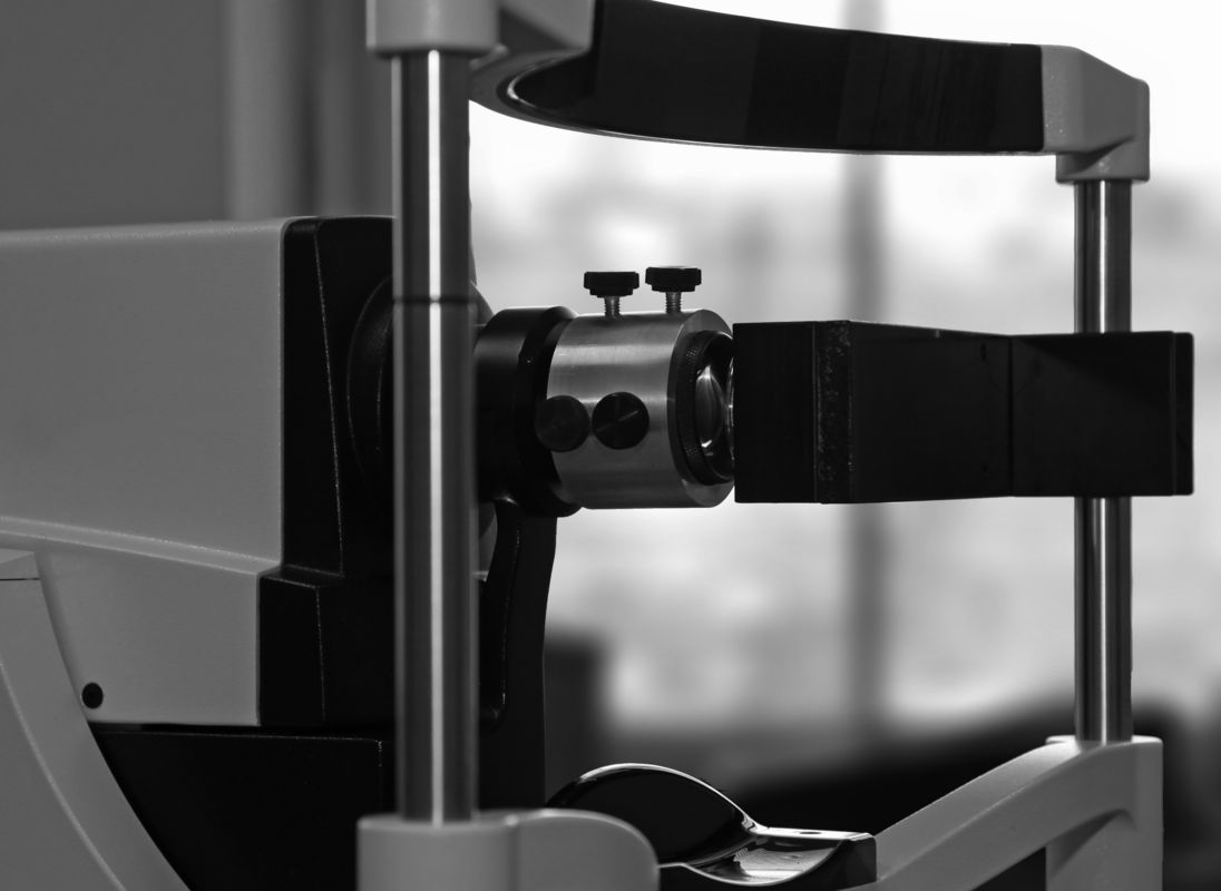
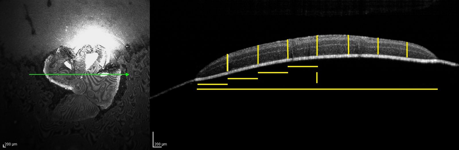
Only a few adjustments had to be made to mimic the in-vivo situation, which the OCT is usually used for. Using a custom-made lens holder (Figure 1 A), an additional lens is mounted in front of the OCT to mimic the lens of the eye. Another custom-made device is used to hold the membrane inserts in an upright position in front of the OCT (Figure 1 B). Two exemplary pictures of the OCT measurement are displayed in Figure 2.
More information:
 |
 |
 |
Retinal degeneration and glaucoma models
 |
 |
 |
 |
Ischemia model
The ex-vivo retinal ischemia model developed in our laboratory combines the advantages of both in-vivo and in-vitro approaches. Retinal explants are nitrogenized in a custom-made ischemic chamber. As only the outer retinal layers have direct access to the culturing medium, there is a limited nutrient supply of the inner retinal layers merely due to diffusion across the retina as in the in-vivo situation. The consequences of retinal ischemia can be studied in a functioning cellular network (the whole retina) and are easily accessible for further investigations.
The use of retinal explants is a suitable way to study the effects of a possible neuroprotective irrigation solution or other treatments after retinal ischemia as this can be directly administered to the retina.
Models of Retinal ganglion cell death
Oxidative damage is a key player in several ocular diseases, including cataracts, uveitis, corneal inflammation, keratitis, age–related macular degeneration and glaucoma. The neurodegenerative influence of local oxidative stress on the retina has been demonstrated. To establish a reliable organ culture model for oxidative stress, degeneration of the retina must be induced by exposure to reactive substances.
Applying three different substances (H2O2, CoCl2 and NMDA), we have developed organ models to study different stages of RGC death. Hydrogen peroxide (H2O2) or Cobalt Chloride (CoCl2), are very strong oxidizers which produce reactive oxygen species (ROS) and led to severe retinal degeneration, whereas NMDA, a commonly applied toxic substance for retinal degeneration, only led to mild apoptotic responses and therefore provides for a model to study the early stages of RGC apoptosis.
Long-term isolated and superfused bovine retina
 |
(in cooperation with Dr. med. Sebastian Müller)
The electroretinogram (ERG) is the standard method for the evaluation of in-vivo photoreceptor function (a-wave) and the function of the inner retina (b-wave). An ex-vivo model that allows for long-term ERG measurements is therefore of high relevance to study the electrophysiology of the eye in a preclinical setting without using laboratory animals. Using bovine retinae of animals from the food industry we have established a long-term ex-vivo culture.
DBA/2J mice
DBA/2J mice are a common natural in-vivo model for pigment dispersion glaucoma. At the age of six to eight months pigment particles from the iris disperse into the anterior chamber leading among other characteristics to an increased IOP and loss of retinal ganglion cells (Figure 5). In several short-term and long-term studies we have monitored the effect of different formulations of glaucoma drugs on the IOP.
 |
Models of optic nerve injury
Models of optic nerve injury (crush or axotomy) can be used to investigate trauma, protection and regeneration of RGCs for studying diseases like glaucoma. They are also very common in neurologic research to mimic traumatic brain injury or neurodegenerative diseases like multiple sclerosis, as the eye is the easily accessible part of the central nervous system.
Ex-vivo Cornea Models
Diseases of the anterior segment of the eye, like dry eye syndrome or corneal infections are generally treated with eye drops. To study those conditions and test new treatment options we have established ex-vivo models of the porcine cornea.
Primary Corneal Epithelia Cells
We have established a method to isolate primary corneal epithelia cells from porcine eyes obtained from abattoirs. With these cells we can conduct a wide range of toxicity tests for novel substances. Cellular viability, apoptosis induction, proliferation rate as well as expression of characteristic markers can be analyzed and give a first hint for the in-vivo situation.
Dry eye
Dry eye syndrome is a chronic condition that involves problems with moistening of the eye´s surface. It is often accompanied by a foreign body sensation in the eye, stinging or reddened eyes, itching and light sensitivity. Due to various causes that are rarely to eliminate completely, constant treatment of the symptoms is required. To test new treatment options in an ex-vivo setting we are establishing cornea organ cultures with an impaired surface. Through surface analysis and measurement of the TEER, as well as molecular markers we can determine the damage and monitor improvement by adding new eye drop formulations.
Infection model to test new efficient treatments
Infections of the eye are generally very painful and can lead to severe impairment of visual functions. The sole effective therapy to combat bacterial eye infections is the repeated daily application of antibiotic eye drops (up to 10-20 times per day). Compliance issues as well as side effects due to highly concentrated active substances result in constant need of new therapy options. To reduce and replace animal experiments we have established an ex-vivo infection model using pseudomonas aeruginosa a common hospital pathogen.