

Neuroretinal Electrophysiology and Imaging
Neuroretinale Elektrophysiologie und Bildgebung
Neuroretinal Electrophysiology and Imaging
Our overarching aim at the Institute for Ophthalmic Research in Tübingen is to join forces to fight the loss of vision. This Junior Research Group focuses on the functional investigation of neuroretina of healthy mice and mouse models of retinal disorders. We apply multidisciplinary strategies for a better understanding of the retinal cells and of the complex network functions.
For vision restoration, the potential of artificial vision devices such as electrical stimulation (retinal electrical implants) and glutamatergic, i.e., retinal transmitter-based implants are presently investigated. Optimal artificial stimulation strategies to replace or support outer retina functions are being pursued to obtain enhanced artificial vision. To achieve such goals, we are supported by highly qualified and dedicated cooperation-partners developing the chip-hardware.
For prevention of vision loss, the potential of electrical stimulation to release neuroprotective factors and that of pharmaceuticals as neuroprotective strategies is investigated. Within this field, we also cooperate with highly qualified and dedicated partners.
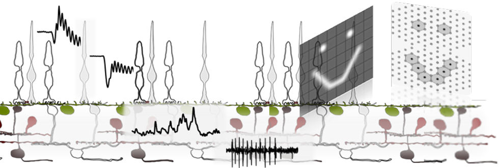
The teams’ multidisciplinary competence in neurophysiology, computer science & engineering allows us to establish and adapt novel and unique experimental setups for neuroretinal electrophysiology by means of extracellular recordings via multi-electrode arrays (MEAs) and simultaneous Calcium-Imaging. The established technique of simultaneous multilayer recording of the retina, allows us to investigate in “sandwich-configuration” the impact of the artificial stimulus on the degenerated outer retina and the corresponding output at the ganglion cell layer. To investigate the impact of a treatment, e.g., pharmaceuticals or genetics, on the functionality of the retinal cells, we have adapted the method of electroretinography (ERG) for retinal explants (acute or cultured) employing MEA recording (mERG). Thereby defined light stimulation protocols allows to discriminate rod and cone photoreceptors as well as cone subtypes. Moreover, the correlated retinal ganglion cell responses are recorded simultaneously, and the functional subtypes are classified. The sensitivity and specificity of these methods also allow us to investigate hIPSCs, such as retinal organoids, together with our cooperation partners.