Dissecting the vertical pathways of the retina
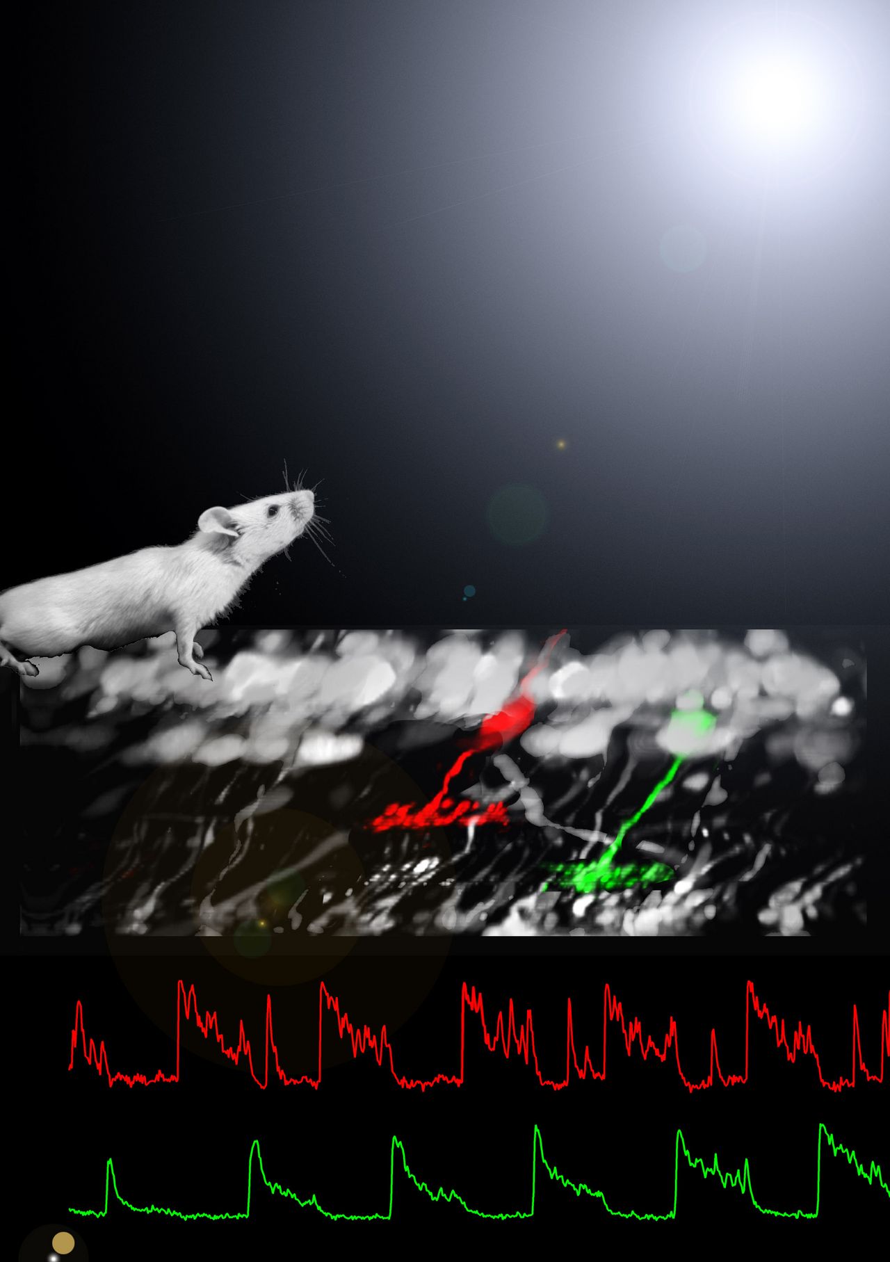
All visual information traverses the retina’s vertical pathway (photoreceptor -> bipolar cell -> retinal ganglion cell), which is split into 10-20 parallel channels. We study visual information processing in all three stages of this pathway towards a better understanding of how and which type of visual information is extracted at different stages of this principal processing chain. Questions addressed here include: What are the specific combinations of ion channels and neurotransmitter receptors that shape functionally distinct bipolar cell pathways in the retina? How do axon terminals of retinal bipolar cells, in interaction with amacrine cells, process information? How is that information integrated in the dendrites of retinal ganglion cells?
Using a combination of visual ecology, calcium imaging in cone pedicles, anatomy and information theory we found that already at the first synapse of visual processing, light response characteristics of the cone array are matched to visual contrast statistics in natural scenes. By imaging of visually evoked synaptic activity in the inner plexiform layer we confirmed the widespread presence of spikes as the main mode of signal transmission by mouse bipolar cells (Baden et al., 2013), in line with recent reports in fish. Moreover, we showed how bipolar cell types in the mouse retina can be clustered by their axonal responses to simple light stimuli. Currently we investigate the intriguing possibility that different terminals belonging to the same bipolar cell might systematically forward different aspects of the visual stimulus to their postsynaptic partners. Moreover, we are interested in how RGCs integrate visual information forwarded from BCs at the level of single synapses.
In analogy to the diversity of signals in bipolar cell axon terminals, we identified different ionotropic glutamate receptors in dendrites of distinct mouse OFF bipolar cell types indicating how the cone signal is funnelled into kinetically distinct parallel OFF channels at the very first synapse in the retina. Furthermore, we found that postsynaptic BK channels contribute to gain control in ON bipolar cell output predominantly at low light levels (Tanimoto et al., 2012).
| Involved Scientists: | Tom Baden, Katrin Franke, Timm Schubert; in collaboration with Silke Haverkamp (MPIB Frankfurt/M), Rob Smith (UPenn), and Rowland Taylor (OHSU). |
|---|---|
| Funding Agency: | Funded by the DFG (CIN-EXC 307) and the Baden-Württemberg-Stiftung. |


